Browse our growing library of real findings from real scans, written by the radiologists who identified them.
A 37-year-old female was referred for orthodontic evaluation. The radiologist noted an enlarged sella turcica in combination with mandibular prognathism. This combination raised suspicion for acromegaly, possibly secondary to a pituitary adenoma. Further diagnostic evaluation including bloodwork was recommended.
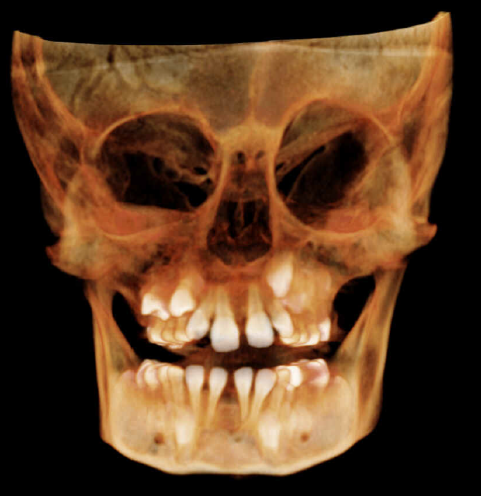
A 10-year-old male presented for an orthodontic evaluation with no reported symptoms. The radiologist identified multiple odontogenic keratocysts and noted findings consistent with possible basal cell nevus syndrome (Gorlin-Goltz syndrome). The number of lesions was unusual, and the upper third molar follicles were noted migrating toward the superior borders of both maxillary sinuses.
A 68-year-old female with no significant medical history was referred for orthodontic evaluation. During full-volume review, the radiologist identified severe erosion and opacification of the right mastoid air cells at the edge of the scan. In the absence of a patient history of surgery, the finding was highly suspicious for a malignant intracranial tumor. The patient was referred for further imaging and evaluation.
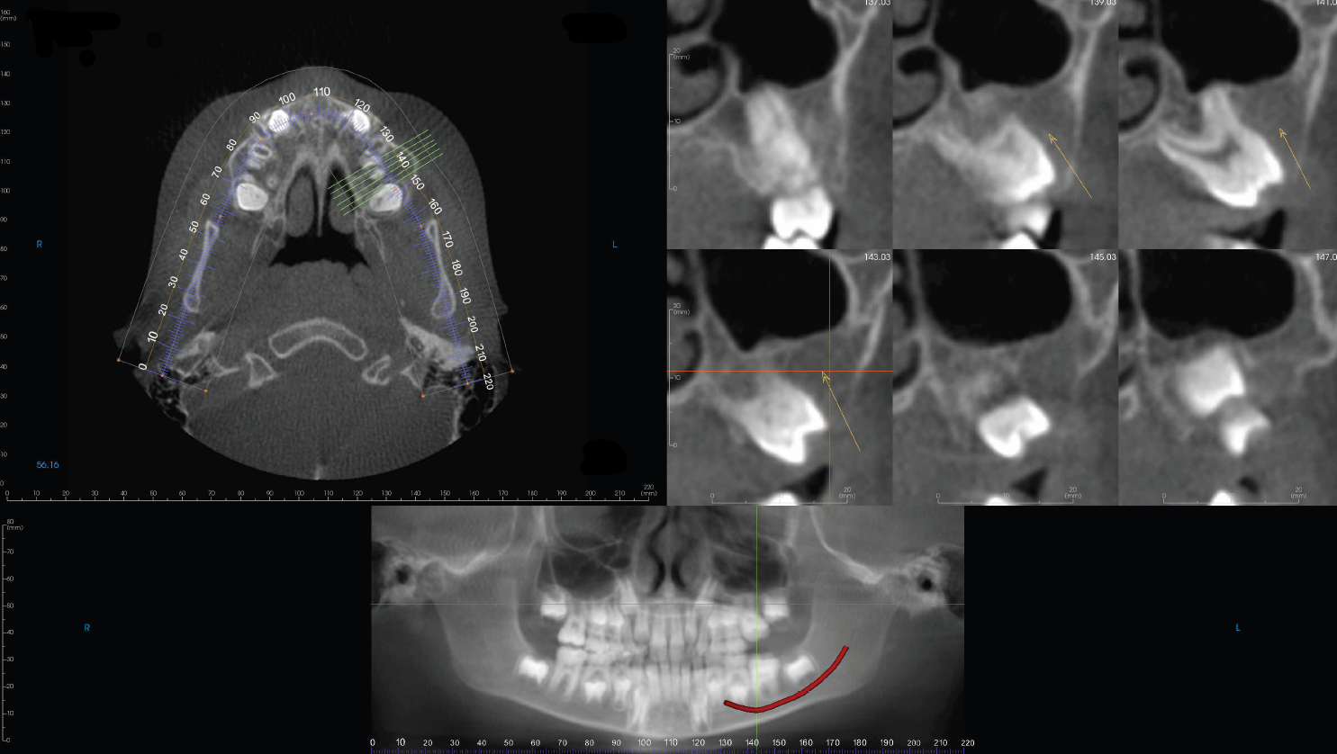
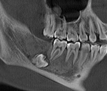
A 59-year-old female was referred for a CBCT scan prior to orthodontic treatment. During full-volume review, the radiologist identified resorption of the lower left canine (#22), with bone growing into the resorbed area suggesting possible ankylosis. The radiologist noted that ankylosed teeth typically do not respond to orthodontic forces, and if undetected, the forces may be transmitted to surrounding teeth, potentially affecting the treatment course and outcome. This finding benefits from specialist review, as it can be subtle and significantly influence orthodontic planning.
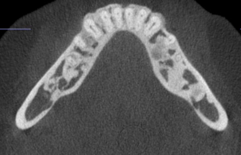
A 24-year-old asymptomatic female was referred for evaluation. The radiologist identified an extreme presentation of what appears as florid cemento-osseous dysplasia, with multiple bilateral patchy to lobulated high to mixed-density foci within the alveolar regions of both jaws. The lesions exhibited a heterogeneous cotton-wool and ground-glass appearance blending with adjacent trabecular bone. No associated cortical expansion, cortical destruction, periosteal reaction, tooth displacement, or root resorption was identified. While typically benign, this condition warrants awareness and monitoring for symptom progression.

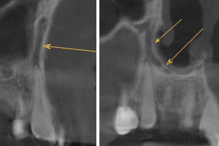
A 67-year-old male was referred for evaluation of possible pathology around tooth #6. The radiologist identified the finding as a canalis sinuosus, an anatomical variation, rather than resorption or pathology. Recognizing this normal variant helped avoid unnecessary workup and provided the clinician with a deeper understanding of anterior maxillary anatomy.
A 72-year-old male currently being investigated for prostate cancer was referred for imaging. The radiologist identified an amorphous radiopacity causing resorption of a tooth, noting that the finding could represent atypical osteosclerosis or a metastatic focus. Given the patient's oncologic history, this differential carries significant clinical weight and warrants further evaluation.
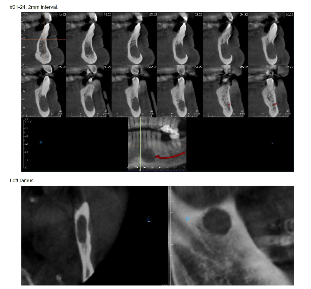
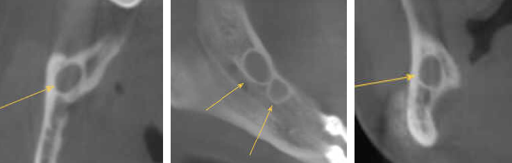
A 44-year-old female with a history of surgical procedures including orthognathic surgery and mandibular plate placement was referred for imaging. The radiologist identified two cysts along the crestal bone within the right mandible. Given the surgical history, these findings were consistent with surgical ciliated cysts. The radiologist noted numerous learning points from this case from both a surgical and clinical standpoint.
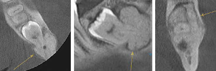
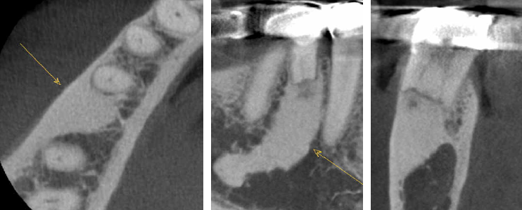
A 16-year-old female was referred for evaluation of a supernumerary tooth in the maxilla. During full-volume review, the radiologist identified a bony fibro-osseous lesion in the mandible with high suspicion of fibrous dysplasia, which appeared to be causing impaction of a separate tooth. This additional finding had not been mentioned in the referral and was likely identified for the first time through dedicated radiologist review.

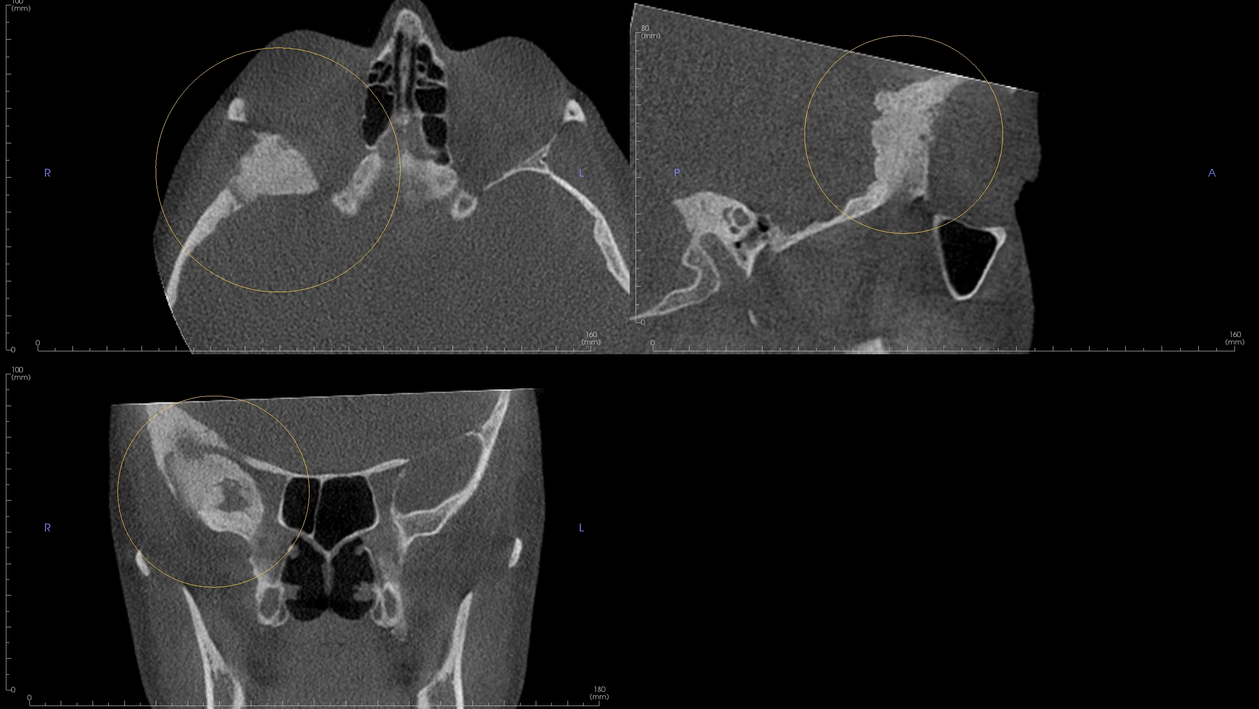
A 41-year-old female with no significant medical history was referred for routine orthodontic imaging. The radiologist identified a sclerotic lesion in the right skull base that may represent reactive hyperostosis from an adjacent brain tumor or fibrous dysplasia. This finding was previously unknown to both the patient and the referring clinician, with no known attributable symptoms. The radiologist noted that while CBCT cannot clearly visualize soft tissue abnormalities, changes to adjacent bone can indicate their presence, warranting further evaluation.

A 46-year-old male presented with an asymmetric, asymptomatic swelling on the right palate. The patient had a history of colon and breast cancer. During full-volume review, the radiologist identified a large fungating growth extending from the nasal cavity into the oropharynx. Given the patient's oncologic history, prompt ENT referral for biopsy was recommended.

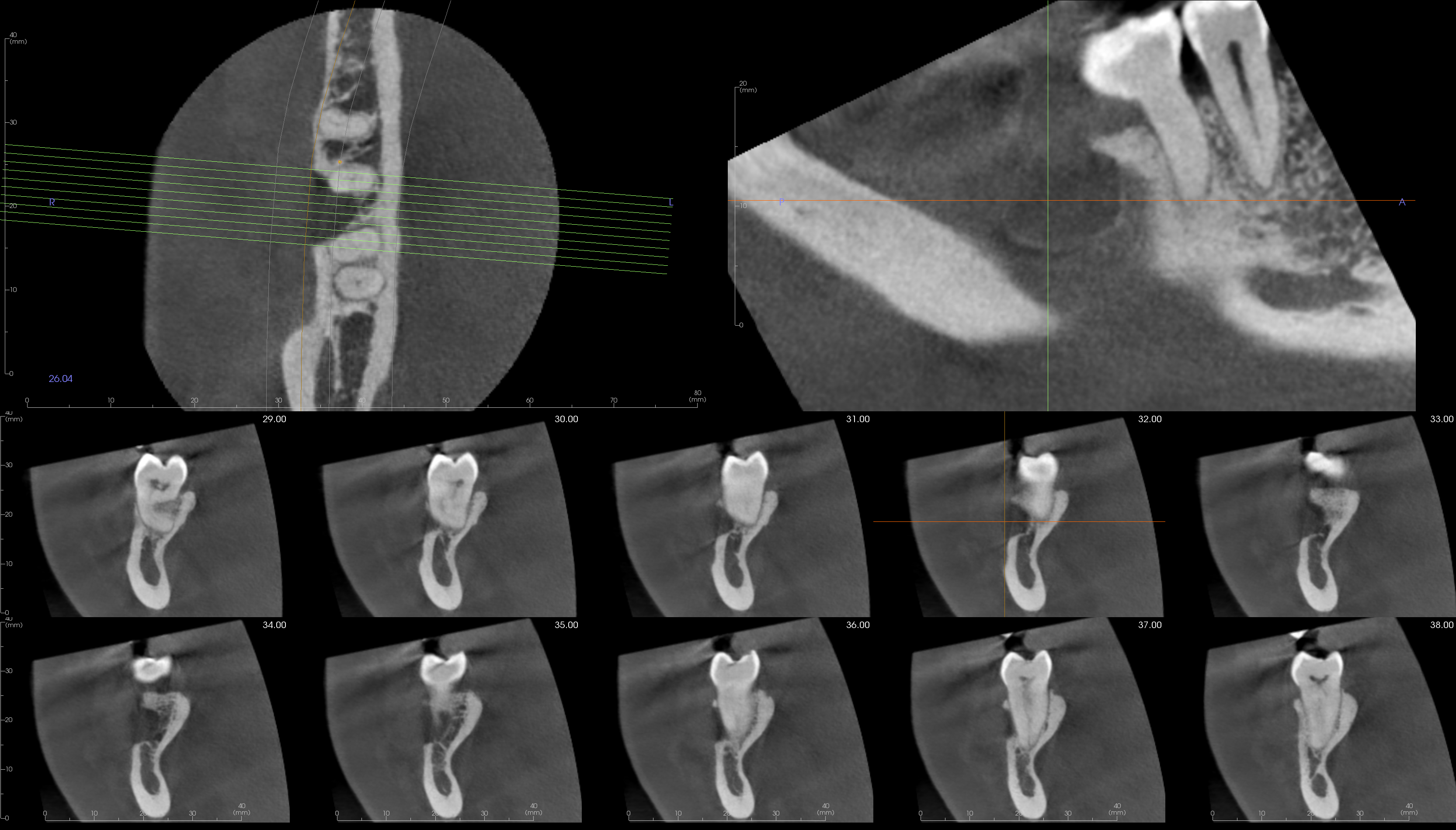
A 43-year-old female was referred with concern over a possible cementoblastoma associated with the left mandibular third molar, encompassing the mandibular canal. The radiologist identified the finding as cemento-osseous dysplasia rather than cementoblastoma, noting the significant difference in treatment implications between the two. The radiologist highlighted this as an excellent teaching case given the complexity of the differential and the range of treatment planning considerations.

A 59-year-old female was referred for evaluation of teeth #6 through #11, all of which had become rapidly mobile, with suspected bone pathology. The radiographic and clinical features were suggestive of a possible malignancy. The finding was ultimately determined to be a very rare but serious fungal infection, which typically affects diabetic patients. This case underscores the value of thorough radiographic evaluation and prompt referral.
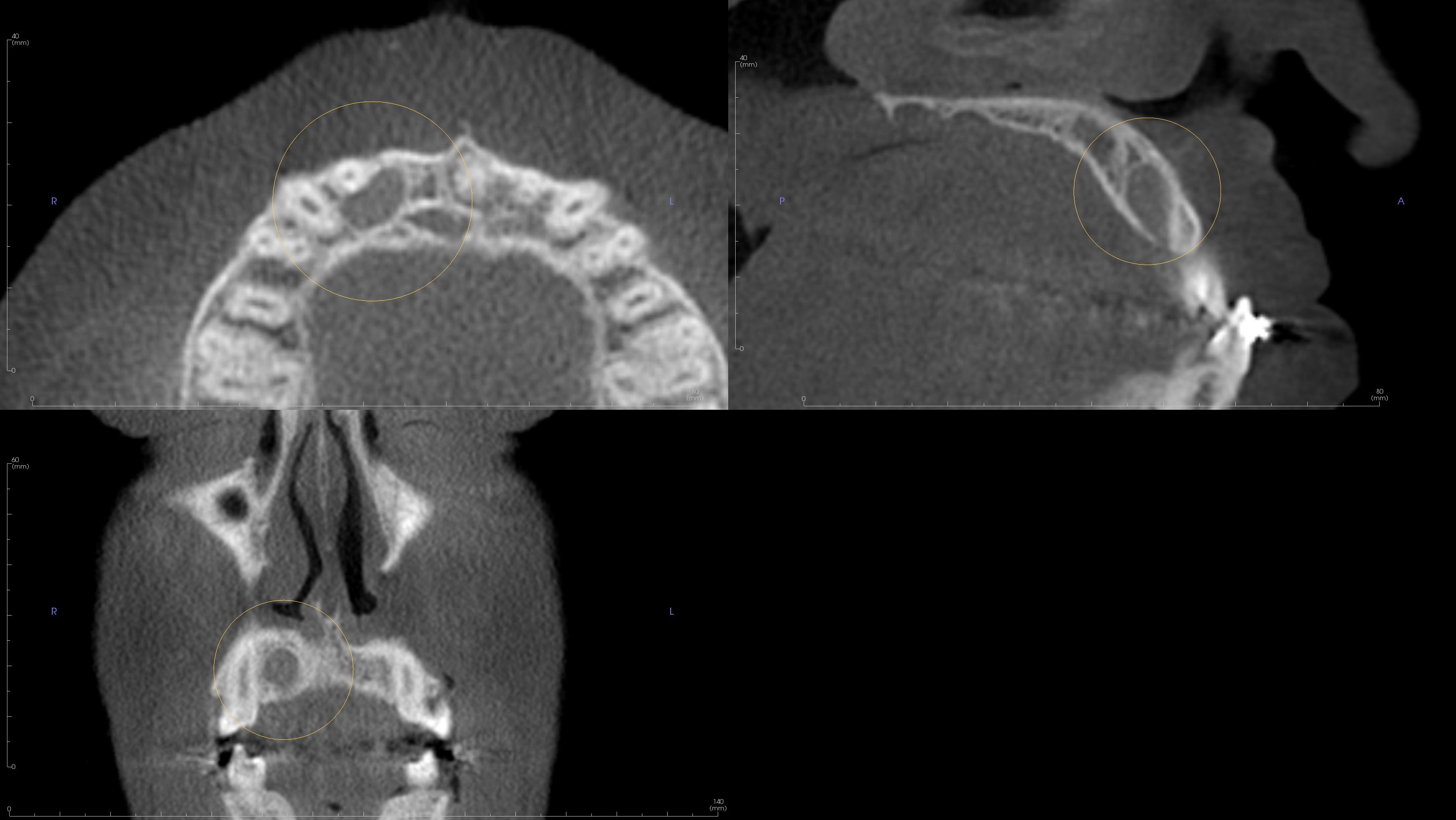
A 9-year-old male was referred for routine pre-orthodontic CBCT assessment. The radiologist identified an arrested mesiodens located within the nasopalatine canal, causing enlargement of the incisive foramen, a very rare anatomic presentation. The radiologist noted that this finding is clinically significant as it would likely be overlooked or misinterpreted on two-dimensional imaging, yet has important implications for orthodontic planning, surgical procedures, and risk of neurovascular involvement. Three-dimensional imaging was essential for accurate localization and characterization of this uncommon finding.
A 76-year-old female without significant medical history had previously undergone excisional biopsy by an oral surgeon for removal of a soft tissue lesion in the left nasal valve, with a reported diagnosis of a nasal cyst. The patient returned to the surgeon a year later with inflammation in the nose. Upon reviewing the scan, the radiologist identified that the lesion had not been completely removed and had in fact increased in size. The surgeon was notified, and the patient underwent further surgery.
A 19-year-old female with no significant medical history was referred for evaluation of an impacted maxillary canine. During full-volume review, the radiologist identified a hypoplastic and opacified right maxillary sinus with an obstructed ostiomeatal unit. The radiographic appearance was suspicious for silent sinus syndrome, and clinical photographs confirmed orbital and facial asymmetry. The radiologist noted that silent sinus syndrome can lead to significant progressive facial asymmetry and may require orbital reconstruction in severe cases. Because the changes develop slowly, they can go undetected for an extended period. The condition is treatable through surgical intervention if the sinus is addressed by an ENT specialist and the ostiomeatal unit becomes patent.
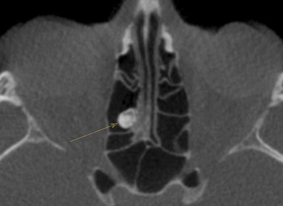
An 83-year-old male with no significant medical history was referred to an oral surgeon due to large maxillary sinuses and the incisive canal observed on a CBCT scan. Upon full-volume review, the radiologist identified a large polypoidal lesion in the left sphenoid sinus extending into the nasal cavity and nasopharynx, suggestive of a large spheno-choanal polyp. Despite the size of the lesion, the patient remained asymptomatic. The radiologist noted that if left untreated, a lesion of this size could cause airway occlusion, breathing difficulty, or extension into the intracranial area. A spheno-choanal polyp of this magnitude is a rare incidental finding in oral and maxillofacial scans, and its detection in an asymptomatic patient underscores the value of full-volume review.
An 18-year-old female with no significant medical history was referred for routine orthodontic evaluation. Upon review, the radiologist identified severely elongated and thickened coronoid processes bilaterally, a condition that can limit mouth opening and mandibular range of motion. The radiologist also noted that one of the zygomas had developed an osteoma, likely from repeated contact with the coronoid process during mouth opening. The radiologist noted that the etiology of limited mouth opening can be difficult to determine and is often attributed to TMJ disorder, but coronoid hyperplasia can be surgically corrected with a very good prognosis. The development of a zygoma osteoma from repeated traumatic contact is a very rare finding.
A 14-year-old female with a history of Treacher Collins syndrome, asthma, tracheostomy dependence, and sinusitis was referred for dental, orthodontic, and surgical treatment planning. The radiologist noted findings consistent with severe Treacher Collins syndrome, demonstrating marked mandibulofacial dysostosis with severe mandibular retrognathia and micrognathia, aplastic mandibular condyles with minimal ramus and glenoid fossa formation, absence or severe hypoplasia of the external ears and external auditory canals, and severe hypoplasia or aplasia of the zygomatic bones and zygomatic arches. The dentition was significantly affected, with severe malpositioning of the mandibular teeth, multiple missing mandibular teeth, and ectopic teeth, including one projecting along the right TMJ articular region.
A 26-year-old female with a history of multiple visits to her physician for symptoms in the area, previously attributed to sinusitis, was referred for evaluation of a radiopaque lesion in the left nostril extending from the palate superiorly with apparent bone expansion on the left palate. The radiologist identified a partially captured aggressive mixed-density expansile lesion centered within the left inferior turbinate and nasal cavity, with osseous remodeling, complete obliteration of the adjacent left nasal airway, and peripheral sunburst-type spiculation along the superior aspect. The differential considerations included juvenile ossifying fibroma, intraosseous hemangioma, osteoblastoma, chondroid neoplasms, and osteosarcoma. Urgent otolaryngology consultation with dedicated contrast-enhanced CT, MRI, and histopathologic evaluation was recommended.
A 55-year-old female was referred by a general dentist for evaluation of a tender facial swelling in the suborbital area adjacent to teeth #6 and #7. The radiologist identified findings on tooth #6 suggestive of apical periodontitis as the likely source of the swelling, along with widespread dental disease including caries and additional apical lesions. During full-volume review, the radiologist also identified multiple punctate soft tissue calcifications in the area of the right parotid gland and possibly the left. The radiologist noted that there is a possible association of these calcifications with other medical conditions, such as sialadenitis or autoimmune conditions. The patient's demographics were noted to be consistent with a profile that warrants further evaluation, as identification of a previously unknown underlying condition could have a significant impact on the patient's overall care and would provide important information for forming dental treatment options, particularly regarding xerostomia and its underlying cause.
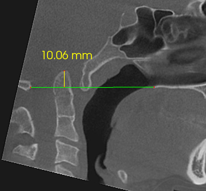
A 72-year-old male had a routine CBCT taken by his dental practice, which identified an incidental finding. The radiologist confirmed a large radiopaque mass within the soft tissues medial to the right posterior mandible, representing an exceptionally large submandibular gland sialolith. The stone was large enough to exert a mass effect on the lingual surface of the right mandible, resulting in mild bone remodeling. The concentric, lamellar appearance was textbook in presentation. Additionally, the radiologist identified diffuse idiopathic skeletal hyperostosis along the anterior cervical spine as a separate incidental finding.
A 9-year-old male with a questionable history of peripheral giant cell granuloma and linear nevus sebaceous syndrome was referred for imaging. The radiologist described this as a highly uncommon syndrome with significant implications, warranting thorough assessment of the craniofacial skeleton and any inflammatory pathology. Imaging revealed cysts, projections within the orbit, and inflammatory processes. The radiologist emphasized the value of this case for understanding the broad craniofacial manifestations of the syndrome.

A 17-year-old female was referred for routine post-orthodontic imaging. The patient was asymptomatic with no significant medical history. The radiologist noted this case as particularly interesting because, although the initial abnormality appeared limited to the sphenoid region, systematic review of the full CBCT volume revealed an extensive skull base lesion with widespread craniofacial involvement extending into the optic canal and vidian canal. Referral for multidisciplinary evaluation and advanced imaging was recommended despite the absence of symptoms, as extensive skull base involvement may have important clinical implications.

A 51-year-old male was referred for orthodontic and TMJ evaluation without significant medical history disclosed. The radiologist identified large post-surgical defects involving the sphenoid sinuses, nasal cavity, nasopharynx, and clivus, along with altered appearance of the left occipital condyle and an asymmetric polypoidal entity in the nasopharynx at the borders of the surgical defect. The radiologist contacted the referring practice, which then confirmed the patient had a history of clivus chordoma removal that had not initially been disclosed. Given the atypical changes near the post-surgical site and the history of cancer, the radiologist referred the patient back to the treating physician for further imaging to rule out residual disease or recurrence.
A 12-year-old female was referred for evaluation of a recurring tender palatal swelling that had been present intermittently for over 12 years. Based on the radiographic and clinical presentations, the referring surgeon was planning surgical intervention. After thorough review, the radiologist identified the finding as consistent with an anatomic variation in the region and contacted the surgeon to discuss the findings. The surgeon was confident in informing the patient that surgery was not necessary at that time.
A 50-year-old African American female with no significant medical history had initially been imaged for radiolucencies in the maxilla and mandible. One site was biopsied, and the histologic report returned cemento-osseous dysplasia, though a cemento-ossifying fibroma could not be ruled out. The patient was asymptomatic and no surgery was performed. After a four-year gap in follow-up, the patient returned with complaints of mandibular enlargement. On the current scan, the lesion had nearly doubled at its largest dimension. Given the prior histopathologic results and the radiographic changes over time, the radiologist made a radiographic determination of cemento-ossifying fibroma. The radiologist noted that this case highlights the role of a radiologist in differentiating between cemento-osseous dysplasia, which is stable and requires no treatment, and a cemento-ossifying fibroma, which has growth potential, since these two entities are histologically identical.
A 75-year-old male was referred for evaluation of spontaneous pain around tooth #2 that had persisted for approximately two years. Upon full-volume review of the CBCT, the radiologist found no significant overt odontogenic pathology on tooth #2 or the adjacent teeth. However, the radiologist identified partial soft tissue opacification with an air-fluid level and frothy secretions in the visualized portion of the right maxillary sinus, along with thickening and sclerosis of the cortical borders and suspected sinus tracts. The findings were consistent with a suspected acute exacerbation of chronic rhinosinusitis of the right maxillary sinus, which required further clinical evaluation and diagnosis. The radiologist noted that the adjacent sinus was a likely source of the patient's pain rather than the dentition, which, if confirmed clinically, would change the treatment approach from dental intervention to sinus management. This case illustrates how full-volume review and 3-D imaging can help differentiate between adjacent structures in the area of chronic pain, which may not be fully appreciated on 2-D imaging alone.
A 24-year-old female with no significant medical history was originally scanned in 2019, when an odontogenic keratocyst was identified and an excisional biopsy was recommended. Follow-up scans submitted in 2020 and 2021 showed no evidence of recurrence. A subsequent follow-up scan in 2023 revealed some unclear findings, and the radiologist recommended additional follow-up imaging in 3 to 6 months. However, when a new scan was submitted in March 2026, 29 months had elapsed since the last radiologist-reviewed scan. The practice also requested comparison with a scan acquired in October 2024 that had not been submitted for radiologist review. Upon review of the current scan, the radiologist identified a large recurrent odontogenic keratocyst that was also present on the 2024 scan and had approximately doubled in size in the intervening period. The patient now presented with pain and swelling. The radiologist noted that the significant growth of the lesion over this period will have a substantial impact on the extent of surgery required and the patient's recovery. This case highlights the value of consistent follow-up imaging and radiologist review for lesions with known recurrence potential.
A 69-year-old male was referred for evaluation of the mandibular bone, especially on the left side. The radiologist identified bilateral irregular, tortuous intraosseous channels within the body and ramus of the mandible, predominantly inferior to the inferior alveolar canals with multiple connections to them. Additional foramina along the lingual cortical surfaces and bilateral lingual cortical thinning were noted without frank cortical destruction or aggressive periosteal reaction, findings most consistent with an intraosseous vascular malformation. The bilateral distribution was particularly unusual, as these lesions are typically unilateral. Advanced medical imaging was recommended to characterize flow dynamics and lesion extent, and invasive dental or surgical procedures should be deferred until the vascular nature is fully characterized, given the potential for dangerous bleeding.
A 37-year-old female with no known significant medical history was referred for a general CBCT evaluation. The referring physician noted the patient had been receiving Botox injections. The radiologist identified a well-defined concavity on the buccal aspect of the left posterior mandible, near the area of attachment of the inferior aspect of the masseter muscle. Upon research, the radiologist found instances of possible bone resorption due to prolonged high-dose Botox injections, where mandibular bone may recede from reduced tension as the masseter muscle is relaxed. The referring clinician confirmed he had never seen a presentation like this in his experience working with TMJ patients, and noted he had discontinued using Botox on patients due to evolving perspectives on the treatment. The clinician expressed interest in reviewing past patients for similar findings and noted this would be a valuable case study.
An approximately 60-year-old female was referred for evaluation of non-spontaneous left-sided facial and infraorbital pain. A periapical radiograph had revealed a periapical radiolucent lesion on tooth #11, and the referring clinician was investigating whether it could be responsible for the pain. A CBCT was acquired for further evaluation, and the referring clinician noted soft tissue thickening in the left maxillary sinus, suspecting a mucus retention pseudocyst. Upon full-volume review, the radiologist determined that the soft tissue thickening was not consistent with a mucus retention pseudocyst. The thickening was causing erosion of the anterolateral and medial sinus walls with extension into the left nasal cavity, as well as demineralization of the left infraorbital canal and left orbital floor. The radiologist noted that these are aggressive findings not associated with benign pseudocysts and recommended the lesion be treated as likely malignant, with referral for further imaging and biopsy. The biopsy confirmed adenoid cystic carcinoma, an aggressive malignancy with a propensity for perineural spread. This case illustrates how soft tissue findings in the sinuses can appear routine but may harbor significant pathology that benefits from specialist radiologist review.
A 15-year-old male was referred for evaluation of a supernumerary tooth in the maxilla and possible supernumerary teeth in the mandible. Upon review, the radiologist identified multiple sclerotic areas in the mandible and maxilla. These areas, in combination with the supernumerary tooth, were suspicious for Gardner syndrome. The patient was subsequently screened and Gardner syndrome was confirmed. As part of the recommended screening process, the patient's immediate family members were also evaluated. During family screening, the patient's mother was found to have gastrointestinal cancer, a known sequela of Gardner syndrome, which was identified at stage 2 and successfully treated with surgery. The patient's mother expressed gratitude to the referring practice, noting that without the radiologist's findings, the cancer would likely have been identified at a more advanced stage. Additionally, the patient is now aware of the genetic condition and can be closely monitored for colorectal cancer going forward. This case illustrates how findings on a routine dental CBCT can have far-reaching implications not only for the patient but for their family members as well.
A 17-year-old male was referred for TMJ evaluation with jaw pain, clicking, and locking. The radiologist identified numerous dense bone islands in both jaws bilaterally, more prominent in the mandible, along with two small osteomas in the right ethmoid air cells. The radiologist noted that the presence of multiple dense bone islands and osteomas strongly suggests Familial Adenomatous Polyposis syndrome, of which Gardner syndrome is a subtype. The referring clinician was advised to correlate with family history of colonic polyps, as these typically develop in the late 30s and carry a risk of malignant transformation over time. Referral to the patient's primary care provider for further diagnostic evaluation was recommended.
A 56-year-old female with no significant medical history presented with jaw joint clicking. The radiologist identified advanced articular remodeling in the right TMJ and degenerative joint disease in the left TMJ with a present reparative phase. Additionally, the radiographic appearance revealed a large loose articular body anterior to the left condylar head, strongly suggestive of secondary synovial chondromatosis in the left TMJ. The radiologist noted that this case raises awareness of the multitude of conditions that can affect the temporomandibular joints, and that it provides a valuable example of secondary synovial chondromatosis with an opportunity to discuss the differences between the primary and secondary forms.
A 75-year-old female with no significant medical history was referred for evaluation of a left TMJ mass identified on panoramic imaging. The radiologist identified a condylar osteochondroma exhibiting corticomedullary-continuous exophytic growth, along with advanced degenerative joint disease and multiple calcified loose bodies within the joint space, findings suggestive of secondary synovial chondromatosis. The coexistence of these entities within a single TMJ is uncommon and presents a diagnostically challenging imaging picture. The radiologist noted that the combination of a benign osseous neoplasm with superimposed intra-articular pathology highlights the value of thorough full-volume review when evaluating TMJ abnormalities.
A 79-year-old male was referred for TMJ examination due to ear and jaw pain. During full-volume review, the radiologist identified a very large soft tissue mass extending from the middle nasal turbinate into the oropharynx, obstructing the patient's airway. The appearance was suggestive of a nasal polyp, though confirmation requires clinical visualization, biopsy, and histopathologic evaluation, as a more aggressive lesion could not be excluded. ENT referral for excision, biopsy, and histopathologic evaluation was strongly recommended. The radiologist noted that the mass was extremely large yet had gone undetected by both the patient and referring dentist, illustrating how CBCT imaging can identify findings beyond the area of clinical concern.
A 61-year-old female was referred for TMJ evaluation. During full-volume review, the radiologist identified severe mucosal thickening in the right maxillary sinus that was displacing the right orbital floor superiorly. The radiologist noted that while the radiographic appearance could be initially interpreted as severe sinusitis, the significant effect on the orbital floor means a pathologic process cannot be completely ruled out. Additionally, even in the case of sinusitis, the degree of orbital floor displacement raises concern that the orbital soft tissue could be affected in rare cases. The radiologist contacted the referring practice as soon as the report was completed and emphasized the need for referral to an ENT specialist to determine appropriate follow-up and rule out any underlying pathology.
A 73-year-old female was referred for TMJ evaluation with no significant medical history provided by the practice. Upon review, the radiologist identified multiple atypical sclerotic and lytic lesions in the TMJs, spine, and skull. Based on the imaging characteristics, the radiologist included metastatic cancer, including breast cancer, in the differential and referred the patient for further workup and imaging. After the report was issued, the practice confirmed that the patient had a long-standing history of breast cancer that had not been included in the referral information. This case confirmed active metastatic lesions in the maxillofacial area and cervical spine, likely altering the patient's treatment protocol.
A 90+ year old male with no significant medical history was referred for TMJ evaluation due to pain and discomfort along the temporomandibular joint. The radiologist identified severe soft tissue asymmetry at the patient's left base of tongue, concerning for possible malignancy. The radiologist called the practice to discuss the finding. The patient was sent to ENT, where squamous cell carcinoma was confirmed.
A 43-year-old female was referred for TMJ evaluation approximately 1.5 months after third molar extractions. The patient had been experiencing persistent symptoms in the left mandible and had consulted multiple specialists, including an ENT, an oral and maxillofacial surgeon, and a TMJ specialist, in an effort to identify the source of the problem. Upon review of the CBCT scan, the radiologist identified radiographic signs of osteomyelitis at the site of tooth #17. Of particular concern, the radiologist noted that the left mandible in the #17 area presented a very high risk for fracture. The radiologist immediately contacted the referring practice after completing the report, recommending that the patient be informed of the radiographic findings and the elevated fracture risk, placed on a soft food diet to minimize stress on the weakened bone, and referred to an oral and maxillofacial surgeon for further management.
A 61-year-old male with pain following third molar extractions was referred for evaluation of the TMJs and extraction sockets. During full-volume review, the radiologist identified a large soft tissue lesion of the right pharynx that was severely displacing and obstructing the patient's airway. Only a few millimeters of the airway remained patent at its narrowest dimension. The lesion was more suggestive of a very large parapharyngeal abscess, though a tumor could not be excluded. Given the severity of the finding and the potential for complete airway occlusion, the radiologist immediately called the referring practice. The case had been read on a Friday afternoon, and the referring doctor contacted the patient directly with instructions to go to urgent care. This case illustrates how full-volume review can identify findings far beyond the area of clinical concern that require immediate action.
A 21-year-old male with no significant medical history presented with pain and swelling on the right masseter with limited range of motion. The radiologist identified severe resorption of the right condyle down to approximately the condylar neck, with periosteal reaction along the condyle and ramus. A comparison scan from three months prior showed an overall normal right condyle, indicating rapid radiographic change. The appearance was most suggestive of septic arthritis, though malignancy could not be ruled out given the rapid destruction. The patient was sent to urgent care and treated with antibiotics, which helped resolve symptoms, supporting the diagnosis. The left condyle showed similar loss of volume that was relatively stable between the two scans. Follow-up imaging at one and three years showed persistent DJD but resolved swelling with no evidence of rapid destruction or periosteal reaction. The radiologist noted that septic arthritis of the TMJ is very rare, and prompt identification helped the patient receive urgent treatment.
A 69-year-old female with chronic TMJ symptoms and a history of multiple craniofacial surgeries was referred for TMD evaluation. The radiologist identified lytic changes in the left skull. Because a scan from 10 years prior was available for comparison, the radiologist could confirm the appearance of the skull had been normal previously. An MRI was recommended. The patient underwent MRI, which confirmed a brain tumor. The patient returned three years later with post-surgical changes showing the tumor had been resected. The radiologist noted that most brain tumors are not visible on CBCT, but this one was causing erosion of the bone, making detection possible. The patient confirmed the lesion had been previously unknown to them.
A 62-year-old female with a history of breast cancer treated with chemotherapy and radiation nine years prior was referred for evaluation of sudden pain in the pre-auricular area and an unspecified sensory deficit in the right mandible. Upon review, the radiologist identified irregular changes of the right condylar head and neck with periosteal reaction, severe sclerosis, and erosive changes. Notably, the right mandibular foramen had an erosive appearance with loss of definition and an obliterated lingula. The radiologist alerted the clinician that the appearance was highly suspicious for breast cancer metastasis. The patient returned to her oncologist and breast cancer metastasis to the mandible was confirmed. Two years later, a follow-up scan was submitted in which the radiologist identified a suspected new breast cancer lesion in the C4 vertebra that was not present on the initial scan. This case demonstrates the importance of radiologist review in identifying metastatic disease in patients with a history of malignancy, even years after treatment, and how follow-up imaging can reveal new sites of disease that may alter the oncologic treatment approach.
A 74-year-old female with a history of cancer was referred for TMJ evaluation by a clinician at a TMJ and sleep practice. The clinician suspected the significant bony changes observed were related to degenerative joint disease (osteoarthritis) and requested a second opinion given the patient's oncologic history. In addition to an osteophyte at the anterior aspect of the condyle, the radiologist identified a large loose ossified body at the posterior aspect of the right condyle. Comparison with prior imaging revealed the ossified body was not present on a 2017 study but had developed by the time of a 2022 scan, and it was associated with malocclusion and a dental midline shift to the left. The radiologist noted that these findings are suggestive of synovial chondromatosis, a rare condition, rather than routine degenerative joint disease. This distinction is clinically significant because the two conditions require different treatment approaches: degenerative joint disease may be managed with splint therapy or observation, while synovial chondromatosis requires surgical removal of the loose bodies and abnormal synovial tissue. If not properly identified and treated, synovial chondromatosis can lead to further joint destruction, occlusal changes, facial asymmetry, and possible extension into the middle cranial fossa.
A 5-year-old male with no significant history was referred for airway and orthodontic evaluation. The radiologist noted opacification of the right ear and mastoids, most suggestive of ear infection. Of note, a prior scan submitted a year earlier showed normal-appearing ears, indicating this was a new development. The patient was referred to ENT and pediatrics for further evaluation.
A 5-year-old patient with no significant medical history was referred for airway evaluation, with plans to be seen by ENT. During full-volume review, the radiologist identified a unilateral parietal calvarial abnormality demonstrating marked thinning with areas of possible full-thickness osseous loss and indistinct inner and outer tables. These imaging features fall into a gray zone between developmental variation and a pathologic process. The radiologist noted that this case underscores the importance of maintaining a broad differential, recognizing the limitations of dental CBCT for calvarial assessment, and prioritizing appropriate referral and advanced imaging when findings are atypical or not confidently benign.
A 14-year-old male with no significant medical history was referred for routine airway evaluation. Upon review, the radiologist identified a large soft tissue lesion occluding the entirety of the nasopharynx. Given the patient's age, the differential included a combination of tonsillar hypertrophy and nasal turbinate hypertrophy or polyps, as well as a nasopharyngeal tumor. The patient was promptly referred to an ENT for further evaluation and management. The radiologist noted that identifying and addressing the lesion will enable the patient to breathe through the nasopharynx again and may help prevent the growth of a potential tumor in a young patient.
A 35-year-old male with no significant medical history was referred for evaluation of the airway, sinuses, and tooth #3. The radiologist identified enlargement and altered trabecular pattern in the left temporal bone region, slightly affecting the left external and middle ear cavities, consistent with fibrous dysplasia. The radiologist noted that while fibrous dysplasia is a benign process, it can cause progressive bone enlargement and narrowing of canals and foramina. The patient should be monitored for left-sided hearing loss, tinnitus, or headaches, with ENT referral warranted if symptoms develop.
A 6-year-old male without significant medical history was referred for orthodontic evaluation. Upon review, the radiologist identified multiple bilateral calcified lesions in the masticator, parapharyngeal, cervical, and submandibular spaces, most suggestive of calcified lymph nodes. One enlarged area along the posterior pharyngeal wall was noted to be causing airway asymmetry. The patient was referred to pediatrics and ENT for further evaluation and to identify the underlying condition, which is typically of infectious origin.
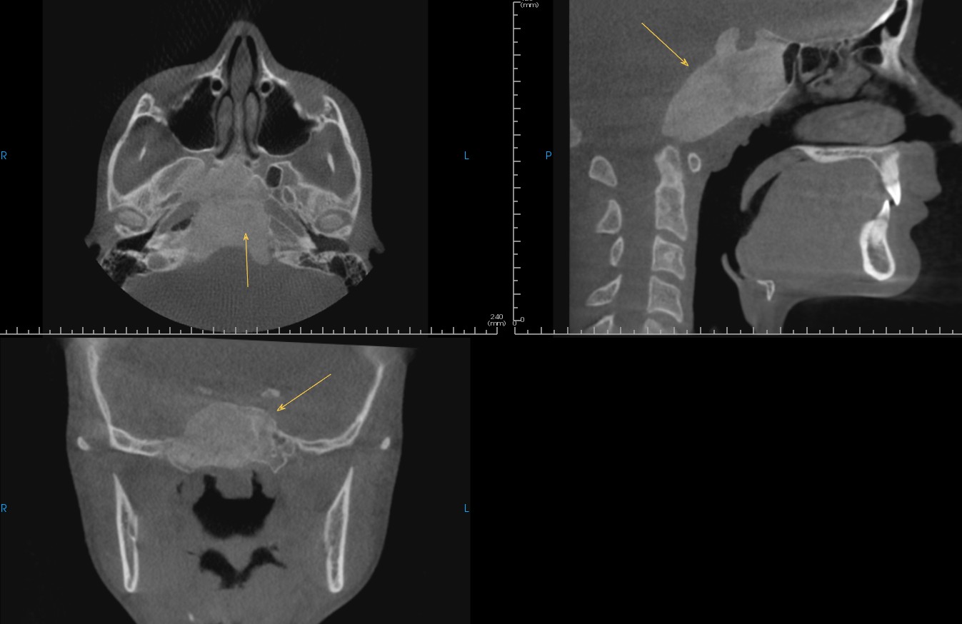
A 60-year-old male with a visible mass behind the soft palate was referred for evaluation of the soft tissue lesion. Upon scan review, the radiologist identified a large polypoidal lesion in the nasal cavity, nasopharynx, and oropharynx that was causing severe airway obstruction. The lesion was considerably larger than what the referring clinician could appreciate clinically. The differential included benign aggressive lesions such as inverted papilloma or a malignancy including squamous cell carcinoma. ENT referral was provided for further treatment.
A 46-year-old male with no medical history provided was referred for an initial airway exam. The radiologist identified a relatively large sphenochoanal polyp obstructing the right posterior nasal choana, a finding that requires surgical intervention to enable proper breathing and prevent further enlargement. The radiologist noted that while antrochoanal polyps are somewhat common, sphenochoanal polyps are more rare. Additionally, a thickened soft palate was noted, which increases risk for obstructive sleep apnea, along with near complete ossification of the right stylohyoid ligament, which may or may not be symptomatic and could be associated with Eagle syndrome.
A 27-year-old male with a history of tongue tie, snoring, TMJ discomfort, and clenching was referred for TMJ and airway analysis prior to lingual functional frenuloplasty. The radiologist identified complete opacification of the right maxillary sinus with reduced volume, inward bowing of the sinus walls, inferior displacement of the orbital floor, and lateralization of the uncinate process, findings consistent with silent sinus syndrome. The left maxillary sinus also showed near-complete opacification with ostiomeatal obstruction, consistent with chronic sinusitis. Silent sinus syndrome is a rare condition where chronic ostial obstruction creates negative intrasinus pressure, causing painless sinus changes that can lead to enophthalmos, hypoglobus, and facial asymmetry. ENT referral was recommended.
A 74-year-old female with no significant medical history was referred for routine airway evaluation prior to fabrication of a mandibular advancement device. Upon review, the radiologist identified severe soft tissue asymmetry in the area of the left palatine tonsils and left base of tongue. The lesion was causing asymmetrical airway narrowing, and although CBCT has insufficient soft tissue contrast for definitive characterization, the appearance was suspicious for squamous cell carcinoma of the left palatine tonsil. The patient, who had been unaware of the lesion, was referred to ENT for further diagnosis and management.
A 7-year-old male was referred for a general airway review. During evaluation, the radiologist identified a foreign body in the right external auditory meatus, partially obstructing the external auditory canal. An ENT referral was recommended for evaluation and removal. The radiologist noted that children often insert small objects in ears and noses that can go unnoticed for extended periods.
A 61-year-old female was referred for evaluation prior to starting oral appliance therapy for obstructive sleep apnea. The radiologist identified a large retropharyngeal lipoma, which was likely contributing to the patient's OSA. The radiologist noted that this is a very rare finding, and that if the lipoma continued to grow without detection, further reduction of the airway would occur.
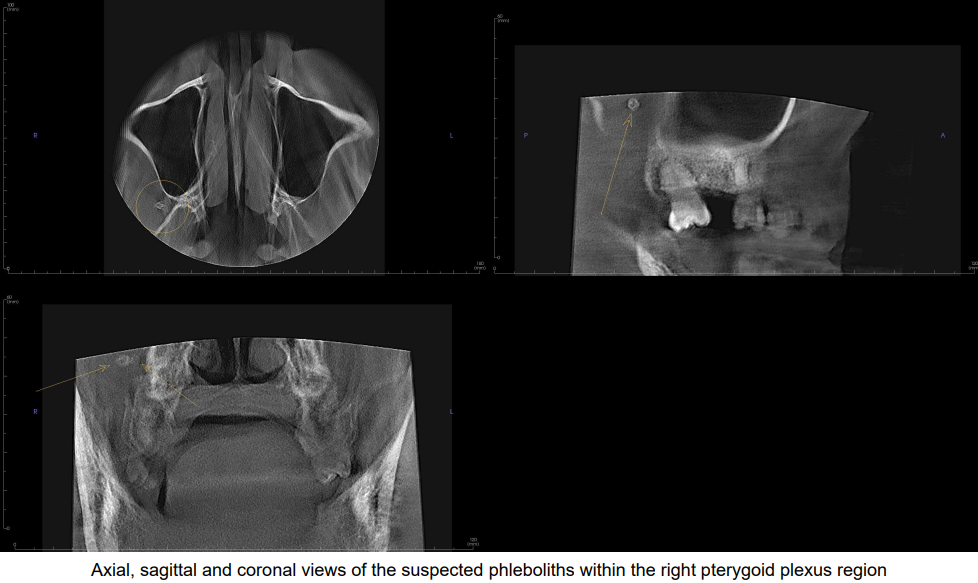
A 70-year-old female with a history of obstructive sleep apnea was referred for airway evaluation. She had undergone a myelogram at approximately 20 years of age. The radiologist identified numerous round radiopacities of very high density scattered throughout the skull base, predominantly on the right side. The radiologist determined these to be persistent oil-based contrast agent from the myelogram performed approximately 50 years prior. The radiologist noted that oil-based contrast agents used in that era can persist indefinitely, unlike modern water-soluble agents, and that myelography has largely been supplanted by MRI, making this an exceptionally uncommon finding.
A 51-year-old male was referred by an airway and sleep practice for a full interpretation of CBCT with no specific clinical notes. During full-volume review, the radiologist identified a severely limited airway with a minimum cross-sectional area of 16.1 mm squared in the oropharyngeal region. At the same time, the condyles were inferiorly and anteriorly positioned while the patient was in maximum intercuspation, and the TMJs presented functional remodeling. The radiographic findings favored a dual bite, a condition in which the condyle positioning and occlusion change due to postural compensation for a small airway. The radiologist noted that dual bite is an orthopedically unstable position for the condyles and cannot be detected except on 3-D imaging with the patient in proper positioning. Any restorative or prosthetic work, or extractions, could allow the condyles to seat further into the fossa, facilitating a change in occlusion that would be difficult for the clinician to manage and potentially significant for the patient. This case highlights the broader effects that a compromised airway can have on the maxillofacial region beyond the airway itself.
A 4-year-old female was referred for evaluation of airway concerns and a narrow palate. During full-volume review, the radiologist identified significant congestion within the middle ears and mastoid air cells, consistent with bilateral otitis media and mastoiditis. The findings prompted a referral to an ENT specialist, who subsequently placed tubes and removed copious amounts of fluid and suppuration. The patient's mother reported that the child's hearing had been significantly impaired, as though she were underwater, but because the child was not experiencing pain or discomfort, the condition would not have been identified without the CBCT review. Following treatment, the patient's hearing improved substantially. The radiologist noted that mastoiditis, if left untreated, can lead to serious complications, making early identification through full-volume review particularly valuable.
An 85-year-old male was referred by an endodontist to evaluate potential root canal failure in #30 and to assess for a fracture. The radiologist identified multiple small fracture lines, confirming a hopeless prognosis for the tooth. The treatment plan was changed from further endodontic treatment to extraction.
A 36-year-old male was referred for endodontic evaluation of tooth #13. During full-volume review, the radiologist also identified a cyst at tooth #16 that was causing bone destruction, tooth displacement, and loss of the sinus floor. Biopsy was recommended. The radiologist noted that identifying and treating this cyst in a younger patient can greatly improve future dental and cosmetic outcomes.
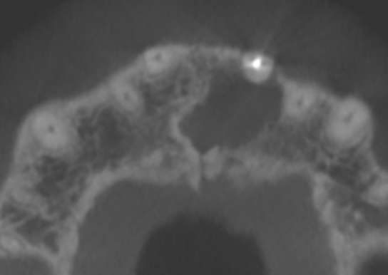
A 20-year-old patient with a history of multiple orthognathic and maxillofacial surgeries was referred for evaluation of root resorption using a small field of view endodontic scan. During review, the radiologist identified a low-density lesion at the scan periphery in the palate, with features suggestive of a surgical ciliated cyst. Referral to an oral and maxillofacial surgeon was recommended. The radiologist noted that this case highlights the importance of reviewing the full extent of even small field of view scans, as incidental findings can be present at the periphery and may be only partially visualized.
A 46-year-old male presented asymptomatic with a history of palatal swelling a few weeks earlier. The radiologist identified a relatively ill-defined radiolucent entity around the apical third of the palatal root of tooth 16, extending to the furcal region with ragged borders, small calcified fragments, loss of the palatal cortical plate, and sclerotic surrounding trabecular bone. While the appearance was suggestive of an inflammatory odontogenic process such as periapical rarefying osteitis, the aggressive imaging characteristics raised concern that a neoplastic process, including a salivary gland malignancy, could not be excluded. Histopathologic evaluation by biopsy was recommended.
A 30-year-old female who first noticed a bump above tooth 8 in 2020 was referred for evaluation. She had been experiencing pressure sensitivity and spontaneous pain. The radiologist identified suspected coalescing persistent periapical inflammatory lesions of teeth 8 and 9, along with an incidental periapical inflammatory lesion on tooth 19. The radiologist noted this case as a strong example of how the OMR's differential diagnosis can influence treatment strategies, with different follow-up approaches suggested depending on whether surgical or non-surgical management is pursued.
A 72-year-old female was referred by a general dentist for review of a periapical low-density lesion on tooth #3. The radiologist identified multiple teeth with apical pathology, but during full-volume review of the large field of view CBCT, also noted soft tissue fullness in the right side of the adenoid tonsil, resulting in asymmetry of the pharyngeal recesses (Fossae of Rosenmuller). The right pharyngeal recess was effaced relative to the left. The radiologist noted that nasopharyngeal cancer on CBCT can present with a similar appearance, as an asymmetrical enlargement of the adenoid tonsil that occludes or effaces the pharyngeal recess. Clinical evaluation by an otolaryngologist was recommended, particularly given that nasopharyngeal carcinoma is most common in older individuals and these imaging findings can precede clinical symptoms. The earliest forms of nasopharyngeal carcinoma are felt to arise in the pharyngeal recess, making this type of finding important to evaluate when included in the field of view.
A 27-year-old female was referred by a general dentist for evaluation of a radiopacity on the distal root of tooth #30, with no reported symptoms. The radiographic and clinical features were suggestive of a dense bone island, a common finding with no clinical significance, though moderate to severe apical external root resorption of the distal root was also present. The radiologist noted that while the severe apical resorption can appear alarming, it is sometimes seen with this condition and does not necessarily indicate cause for concern on its own. However, external cervical resorption was also identified on the same tooth, which means periapical sclerosing osteitis should be considered in the differential. The radiologist noted that the radiographic density, borders, and extension of the apical lesion, along with the absence of symptoms, are more characteristic of dense bone island, but action may still be needed to address the resorptive process.
A 78-year-old male presented with a radiolucency associated with the left maxillary central incisor that appeared to erode the nasopalatine canal. The radiologist noted this as a valuable teaching case, as the ambiguous nature of the finding raises the question of whether the radiolucency originates from the tooth or from the nasopalatine canal itself. Biopsy was recommended to determine the definitive origin, illustrating that imaging does not always provide a clear-cut answer.

A 60-year-old male was referred from an endodontic practice with clinically significant palatal swelling and soft tissue ulceration with rolled borders, despite vital teeth in the region of interest. The radiologist identified a lesion causing considerable expansion of the dentoalveolar region in multiple directions. The appearance was suggestive of a benign neoplasm, most likely an ameloblastoma, with implications for multiple adjacent anatomical structures. Given that surgical treatment would involve wide margin of resection, the patient would likely require advanced prosthodontics including a custom obturator device.
A 61-year-old male with no significant medical history was referred for follow-up of endodontic treatment and comparison of an apical lesion around #2 with prior scans. The radiologist identified extensive emphysema in the right buccal soft tissues and zygoma, possibly secondary to endodontic treatment. The referring clinician had been unaware of the emphysema. The radiologist noted that this finding may be confused with a dental abscess and can go undetected, and that uncontrolled emphysema can be very painful and potentially dangerous if it spreads.
A 74-year-old female was referred by a periodontist after an endodontist noted a periapical radiolucency at the apex of tooth #9. The referring clinician observed that the nasopalatine canal appeared enlarged and wanted to differentiate between a nasopalatine duct cyst and an apical lesion originating from #9, as the two differential diagnoses require different treatment approaches. Based on the shape of the lesion and detailed evaluation using radiology software to view the pathology from multiple angles, the radiologist determined the primary differential to be a persistent periapical inflammatory lesion associated with #9. The radiologist noted that this case highlights the value of an oral and maxillofacial radiologist in identifying nuances that differentiate one diagnosis from another, providing a perspective that multiple specialists had been unable to determine.
A 39-year-old male was referred by a general dentist for evaluation of pain in the mandibular anterior teeth. The radiographic findings were suggestive of intermediate-stage periapical cemento-osseous dysplasia on teeth #23 through #27, with suspected perforations in the adjacent cortices. The radiologist noted that with these findings, the possibility of secondary infection should be considered. When this fibro-osseous lesion becomes exposed to the oral environment, secondary infection with concomitant sequestration of the internal dense mass may occur, often leading to intense pain. This changes the clinical approach from treating it as a lesion that generally does not require intervention to one requiring prompt surgical removal of infected tissue by an oral surgeon. The radiologist noted that many general dentists may not be fully aware of the potential complications associated with this lesion, since it typically does not require treatment, and this lack of awareness may lead to delayed action.
An 87-year-old female presented with a large firm mass under her chin in the midline. A medical ultrasound had been inconclusive, and the physician requested a dental evaluation. The patient had prior root canal therapy on teeth #26 and #27, which were asymptomatic, and the referring dentist noted associated apical lesions with some apparent bone fill. The dentist questioned whether there was evidence of incomplete osseous healing from the previous root canal therapy contributing to the mass. Upon full-volume review, the radiologist determined that both apical lesions showed no apparent radiographic signs of osseous healing, with adjacent perforation of the facial cortex and adjacent soft tissue swelling both facially and inferiorly. While CBCT is not ideal for soft tissue assessment, the radiologist noted that these findings suggest cellulitis, phlegmon, or abscess, which are varying degrees of soft tissue inflammation requiring prompt treatment to prevent further spread of infection through the soft tissues. This case demonstrates the oral radiologist's ability to bridge the gap between the general dentist's and physician's expertise and to accurately guide the dentist's radiological interpretation when uncertain, facilitating quick and decisive treatment.
An 80-year-old male was referred by an endodontist for evaluation of what appeared to be a large endodontic lesion. Upon review, the radiologist identified clinical features more consistent with malignancy rather than a simple dental infection. Consultation with the referring office confirmed the patient had been found to have multiple myeloma. The radiologist noted that the distinction between an endodontic lesion and a malignant process significantly changes the course of treatment.
A 66-year-old female with a medical history of aneurysm surgery was referred for implant evaluation. During review, the radiologist identified a well-defined, high-attenuating, circular structure in the region of the foramen magnum, possibly associated with the right vertebral artery. The finding was noted to be consistent with aneurysm embolization material, specifically a coil mass, correlating with the patient's reported surgical history.
A 58-year-old female was referred for implant site evaluation. The radiologist incidentally identified a carotid artery calcification, though not in the typical location at the bifurcation. The calcification was positioned more toward the midline, suggestive of a retropharyngeal carotid artery calcification, a finding the radiologist noted is uncommon. Its atypical location could have been confused with a different anatomical structure such as the hyoid or thyroid, potentially leading to insufficient follow-up.
A 49-year-old male was referred for implant evaluation at the left maxillary #14 site. During full-volume review, the radiologist identified that the right maxillary sinus was not only hypoplastic but that the anterior and lateral walls were caved in, with the orbital floor bowed inferiorly. The sinus was essentially clear. The radiologist suspected silent sinus syndrome and recommended clinical correlation for signs of hypoglobus or enophthalmos. The ostiomeatal complex was not fully captured in the scan, and further evaluation was recommended.
A 37-year-old female with a history of pain in the left ramus area after a traumatic event was referred for evaluation of possible fractures or other causes. The radiologist identified the implant apex at site 18 extending into the superior half of the inferior alveolar canal with associated canal narrowing, despite normal crestal peri-implant bone levels. Implant extension into the inferior alveolar canal carries an increased incidence of neurosensory disturbance, including paresthesia, dysesthesia, or chronic neuropathic pain. Neurosensory assessment was recommended, along with counseling the patient regarding the elevated risk of developing symptoms and the need for close monitoring.
A 73-year-old female experiencing discomfort in the region of implant 19 was referred for evaluation. The radiologist identified a cosmetic implant along the facial surface of the mandibular body extending from anterior to the left mental foramen to approximately the right retromolar area. The implant demonstrated asymmetric positioning and mild cortical bone erosion in the anterior mandible. Its position inferior to the right mental foramen was noted as clinically significant, as external compression of the mental nerve can cause lower lip and chin paresthesia. Correlation with clinical signs, symptoms, surgical history, and comparison with prior imaging were recommended to determine whether the implant had migrated from its original position.
A 70-year-old male with depression, HIV, and malignant tumors of the colon and prostate was referred by a general dentist planning all-on-x procedures. The radiologist identified several periapical inflammatory lesions and lesions of cemento-osseous dysplasia. However, an additional poorly defined intra-osseous low-density lesion was noted in an edentulous site. While this could represent a residual inflammatory lesion or cemento-osseous dysplasia, the radiologist noted that an aggressive lesion of non-inflammatory etiology, specifically metastatic disease, needs to be ruled out given the patient's history of colon and prostate malignancy. The radiologist noted that this type of finding may be easily overlooked when several other inflammatory lesions are present, since malignant and inflammatory lesions share radiographic features, and that the prevalence of malignancy and metastasis is increased in older patients with HIV.
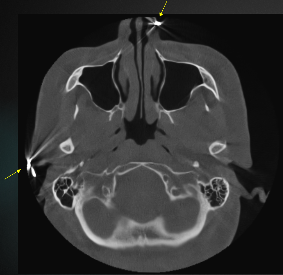
A 57-year-old female was referred for implant planning for reconstruction of terminal dentition with implant-supported dentures. During review, the radiologist identified beam hardening artifact caused by multiple pieces of jewelry, including earrings and a nose piercing, that had not been removed prior to image capture. The radiologist noted that one earring was positioned just outside the field of view yet still produced artifact, illustrating that objects in the path of the X-ray beam can degrade image quality even when not within the scanned volume. Removing all jewelry before CBCT capture is a straightforward step that can significantly improve image quality for interpretation.

A 65-year-old male was referred for implant placement evaluation. During full-volume review, the radiologist identified calcifications within the vertebral arteries. This finding typically indicates significant plaque burden and suggests other vascular areas may also be affected. Referral to the patient's primary care provider was recommended for evaluation of atherosclerosis. The radiologist noted that while carotid calcifications are more commonly identified, vertebral artery calcifications should not be overlooked.
A 74-year-old male was referred for implant placement in the area of the right maxillary first molar. The radiologist identified a surgical ciliated cyst in the area of a previous sinus lift. This type of cyst can develop when instruments scrape respiratory epithelium during surgery and that tissue contacts another area of the jaw. The radiologist noted this finding has significant implications, as the cyst requires management before implant placement can proceed.
A 48-year-old male with multiple missing, mobile, and supra-erupted teeth, along with moderate to advanced periodontal disease, was referred with all-on-x procedures planned. The radiologist identified well-defined destruction of much of the alveolar process around the maxillary and mandibular left posterior teeth, with scooped-out and floating-tooth appearances. While perio-endo inflammatory lesions are likely contributors, the radiologist noted that Langerhans Cell Histiocytosis, such as eosinophilic granuloma, should be ruled out with biopsy, especially since several affected teeth lacked coronal destruction or restoration. The radiologist noted that this condition is uncommon and would require surgical treatment and possible radiation therapy, chemotherapy, or intralesional corticosteroids before implant treatment could be considered. Radiographically, this could be fairly easily mistaken for aggressive periodontal disease, potentially leading to inappropriate treatment.
A 71-year-old female with no significant medical history was referred for evaluation of dental implant #14. During full-volume review, the radiologist identified a linear radiopaque entity in the base of the tongue anterior to the vallecula on the right side. The radiologist noted that the base of the tongue is a common site for impaction of sharp food objects such as fish bones, and that migration, infection, and injury to nearby structures are ongoing risks. As the referring clinician was an oral surgeon, clinical evaluation was recommended.
A 64-year-old male was referred for implant placement evaluation at the #7 site. During full-volume review, the radiologist identified expansion of the middle cranial fossa. The radiologist contacted the referring practice to discuss the finding, and the clinician noted they had not been aware that the brain was included in the field of view. The finding was determined to be a slow-growing, benign brain tumor. This case illustrates the value of comprehensive CBCT review beyond the area of clinical interest.
A 46-year-old female was referred by a general dentist for evaluation of the right maxillary edentulous area for implant placement. The patient had a history of multiple supernumerary teeth in the area when she was younger, along with a prior surgery in which she reported that "cement" was placed. During full-volume review, the radiologist identified a well-localized, high-density, mildly expansile lesion with a largely dense, amorphous bone appearance, but also areas of dense ground glass pattern and cotton-wool pattern. Vertically-oriented, thick trabeculae were also appreciated. The radiographic findings were suggestive of fibrous dysplasia versus segmental odontomaxillary dysplasia, though bone grafting could not be ruled out in a portion of the lesion. The radiologist noted that segmental odontomaxillary dysplasia is a rare condition that was considered due to the vertically-oriented trabeculae combined with the observed and reported tooth abnormalities, including external root resorption and partly calcified pulp spaces. These pathologic conditions are characterized by abnormal bone, and the radiographically dense appearance likely suggests decreased blood supply. The radiologist recommended caution with any surgical procedures in the area, as the findings would likely change the referring dentist's treatment plan. Comparison with prior imaging and consideration of surgical history was also recommended to further delineate between the two conditions.
A 41-year-old male, new to the referring practice with no previous medical history on file, came in for a routine exam and cleaning. On review of the scan, the radiologist identified extensive sinonasal changes including palatal cleft, septal perforation, turbinate loss, absent ethmoid sinus floor, discontinuous maxillary sinus walls with possible sequestra, collapse of nasal alae, enlarged palatine canals/foramina, and cleft palate. These findings may be suggestive of conditions such as Granulomatosis with polyangiitis (Wegener's granulomatosis), osteonecrosis, or chronic invasive fungal sinusitis. An expedited referral to ENT was recommended. The radiologist noted this degree of pathology is an uncommon finding, particularly in a patient not currently undergoing treatment, and expressed appreciation for the opportunity to bring these changes to the patient's attention.
A 73-year-old female with TMJ disorder and airway congestion was submitted for sinus, airway, and TMJ evaluation. While reviewing the full scan, the radiologist identified a hyperdensity within the soft tissues of the brain, indicative of either a brain bleed or malignancy. The radiologist called the practice first thing in the morning and spoke with the provider. The patient was the provider's mother. An MRI was recommended immediately. The patient had an MRI that same day, which confirmed the intracranial entity to be a meningioma. The patient had no specific symptoms that would have prompted further imaging. The radiologist noted this was a completely incidental finding that, if undetected, could have gone unnoticed for an extended period.
No cases in this view yet. New cases are added as our radiologists submit them.
All cases are de-identified. Details are limited to what the submitting radiologist reported; findings are identified on imaging and confirmed through clinical correlation by the treating team.
Learn which of your own cases may benefit most from a second set of eyes in our eBook, The Traffic Light Protocol for OMR Referral.
Download the free eBookRegistering is easy, free, and secure.
As low as $89 per report, authored by an oral and maxillofacial radiologist. No subscriptions, no minimums.