Every reader here is an oral and maxillofacial radiologist, so your treatment plan rests on full-volume specialist interpretation.
Get a structured, comprehensive interpretation of the full imaged volume: findings, impressions, and follow-up guidance written to support treatment planning.
 Radiology Report
Implant evaluation for site #3 in the posterior maxilla with attention to sinus anatomy and ridge dimensions.
No radiographic signs of bone pathology were noted at site #3. A septation was identified in the sinus apical to the site. The left ostiomeatal complex was not patent, and moderate to severe mucosal thickening was noted in the left maxillary sinus. Subantral height measured approximately 4.21 mm.
Calcified atheromas were noted in the internal carotid arteries intracranially within the circle of Willis area.
Site #3 showed no bone pathology, though the sinus septation should inform implant planning. Sinusitis cannot be ruled out from the left maxillary sinus given the mucosal thickening and non-patent ostiomeatal complex. Medical history evaluation is suggested to assess cardiovascular risk.
BeamReaders was co-founded in 2007 around one oral and maxillofacial radiologist, Dr. David Hatcher, who saw a gap between what dental practices needed and what traditional radiology delivery could support. What began as one radiologist reading scans has grown into a specialist institution with shared standards, mentoring, and peer collaboration.
BeamReaders serves everyone from a solo practitioner to a DSO running hundreds of locations, with the technology platform and clinical infrastructure built around how practices operate.
Full-volume review and structured reporting that supports treatment planning.
We recruit, mentor, and continuously develop oral and maxillofacial radiologists.
BeamReaders' purpose-built platform, VoxDental, enables you to submit your first case, or your thousandth, in minutes.
Radiologists and practices work together through clear, documented communication.
Our collective case experience informs practical frameworks like the Traffic Light Protocol, helping teams categorize cases and prioritize referrals with consistency.
Download the Free eBookIn our experience, these carry the greatest diagnostic complexity and risk of relevant incidental findings. Most practices benefit from specialist review.
Elevated risk, especially when outside your training, experience, and comfort level. If in doubt, referral is recommended.
Straightforward, routine cases. If you self-read, review the entire volume systematically and document it. The eBook offers tips on how to do this.
A 67-year-old male was referred for evaluation of possible pathology around tooth #6. The radiologist identified the finding as a canalis sinuosus, an anatomical variation, rather than resorption or pathology. Recognizing this normal variant may have helped avoid unnecessary workup and provided the clinician with a deeper understanding of anterior maxillary anatomy.
A 90+ year old male with no significant medical history was referred for TMJ evaluation due to pain and discomfort along the temporomandibular joint. The radiologist identified severe soft tissue asymmetry at the patient's left base of tongue, concerning for possible malignancy. The radiologist called the practice to discuss the finding, and on that recommendation the practice referred the patient to ENT, where squamous cell carcinoma was confirmed.
A 61-year-old female was referred for evaluation prior to starting oral appliance therapy for obstructive sleep apnea. The radiologist identified a large retropharyngeal lipoma, which was likely contributing to the patient's OSA. The radiologist noted that this is a very rare finding, and that if the lipoma continued to grow without detection, further reduction of the airway would occur.
An 80-year-old male was referred by an endodontist for evaluation of what appeared to be a large endodontic lesion. Upon review, the radiologist identified radiographic features more consistent with malignancy rather than a simple dental infection.
Consultation with the referring office confirmed the patient had been found to have multiple myeloma.
The radiologist noted that the distinction between an endodontic lesion and a malignant process significantly changes the course of treatment.
A 66-year-old female with a medical history of aneurysm surgery was referred for implant evaluation. During review, the radiologist identified a well-defined, high-attenuating, circular structure in the region of the foramen magnum, possibly associated with the right vertebral artery. The finding was noted to be consistent with aneurysm embolization material, specifically a coil mass, correlating with the patient's reported surgical history.
Every case is interpreted, reviewed, and documented by an oral and maxillofacial radiologist. Follow up questions go to them directly via Rad Chats in VoxDental.
This enables BeamReaders to coexist with the AI screening software many practices already use, as screening and interpretation are different jobs. Your radiologist reads the full volume in your patient's clinical context, addresses your clinical question and study purpose, and signs the report.
I wouldn't feel safe taking a CBCT without BeamReaders looking at it with me.
Set up your free VoxDental account. No subscriptions, fees, or minimums.
See How It WorksAdd your scan files and clinical context in one secure submission workflow.
Tips on Sending Your CasesAccess reports, follow up with your radiologist, and collaborate with the full care team.
Submit your first casePractical training and ongoing learning, built and delivered by the radiologists who read your cases.
Self-paced CE curriculum on CBCT fundamentals and more.
Small-group classes led by preeminent OMRs in the field.
Direct guidance tailored to your clinical focus and workflow.
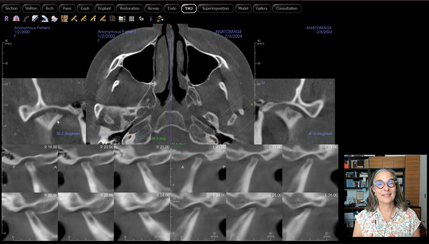
A quick, insightful recap of our recent session on reading a cone beam CT by Dr. Dania Tamimi.

Dr. Anna Csillag on a lifelong career of oral radiology and finding where she belonged.

This article is based on a webinar led by Dr. Srinitha Singanamala and Dr. Anne Dorsey, Oral and Maxillofacial Radiologists at BeamReaders. In the session, they walked through how to turn your CBCT submission into the most useful report possible, from the moment you decide to refer to the moment the report lands back in your inbox. The basis is simple: great information in, great report out.
In just a few minutes, go from new account to requested report.
As low as $89 per report, authored by an oral and maxillofacial radiologist. No subscriptions, no minimums.