20-year-old male
A 20-year-old male with no significant medical history was referred by his orthodontist for TMJ evaluation and orthodontic planning prior to beginning treatment. During review, the radiologist identified a mixed-density lesion with faint internal calcifications in the right anterior maxilla, with radiographic features most suggestive of a benign odontogenic tumor or cyst such as an adenomatoid odontogenic tumor or calcifying odontogenic cyst. The finding was incidental and the patient was asymptomatic.
The radiologist noted that lesions with internal calcifications are relatively rare in the anterior maxilla, and that left unaddressed, a lesion of this kind would likely lead to significant further complications, including more difficult and prolonged orthodontic treatment and potential loss of regional dentition. The radiologist recommended that the lesion be addressed before orthodontic treatment begins.
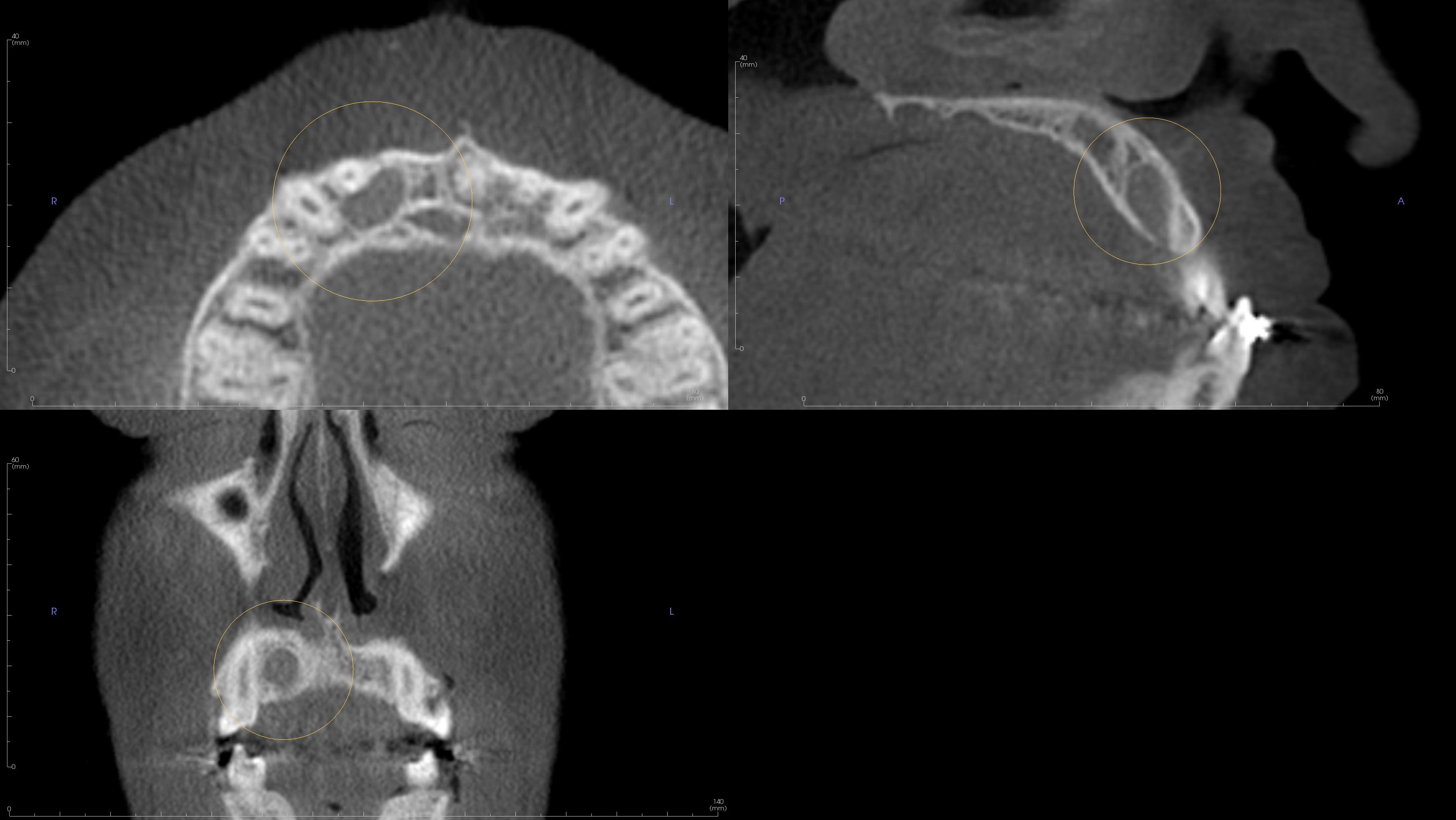
De-identified scan detail.
Registering is easy, free, and secure.
As low as $89 per report, authored by an oral and maxillofacial radiologist. No subscriptions, no minimums.
