74-year-old male
A 74-year-old male was referred for a general review of his CBCT scan with no additional clinical information provided. The scan had originally been taken to evaluate a draining fistula on tooth #30, and the large radiolucency in the lower anterior region was an incidental finding.
During comprehensive evaluation, the radiologist identified multiple low-density areas, including a second lesion in the left ramus that was not mentioned in the referral. The presence of multiple lesions raised suspicion for a systemic abnormality, prompting the radiologist to contact the referring clinic for medical history. It was then learned that the patient had a history of plasmacytoma, first identified several years prior, which had returned on his pelvic bone near a hip prosthesis. The patient had recently completed two sessions of proton radiation therapy and was awaiting a PET scan before starting chemotherapy.
This case illustrates how obtaining medical history, combined with thorough scan review, can connect seemingly unrelated findings to a known systemic condition and support the clinical team in coordinating the patient's care.
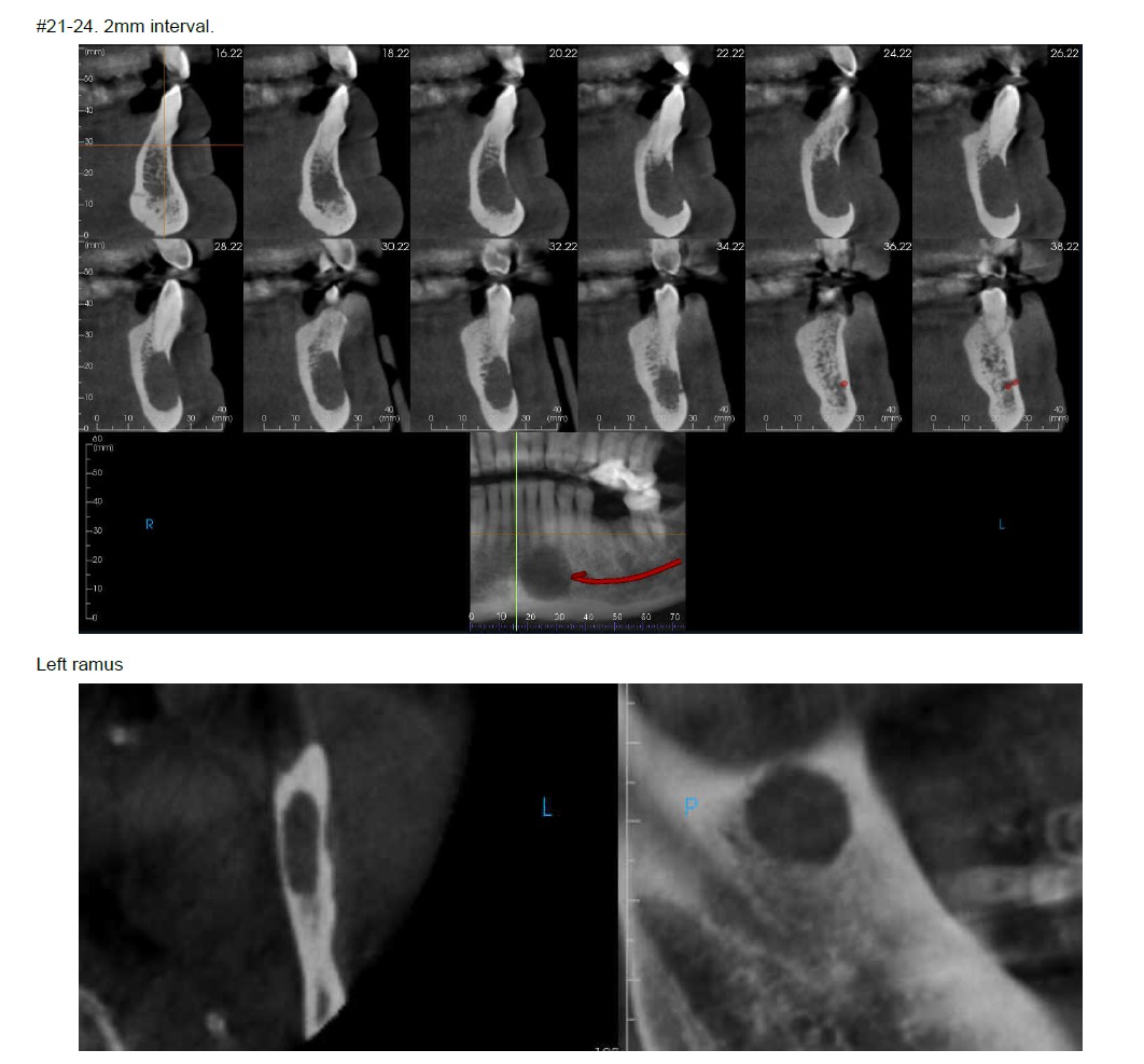
De-identified scan detail.
Registering is easy, free, and secure.
As low as $89 per report, authored by an oral and maxillofacial radiologist. No subscriptions, no minimums.
