41-year-old female
A 41-year-old female with no significant medical history was referred for routine orthodontic imaging. The radiologist identified a sclerotic lesion in the right skull base that may represent reactive hyperostosis from an adjacent brain tumor or fibrous dysplasia.
This finding was previously unknown to both the patient and the referring clinician, with no known attributable symptoms. The radiologist noted that while CBCT cannot clearly visualize soft tissue abnormalities, changes to adjacent bone can indicate their presence, warranting further evaluation.
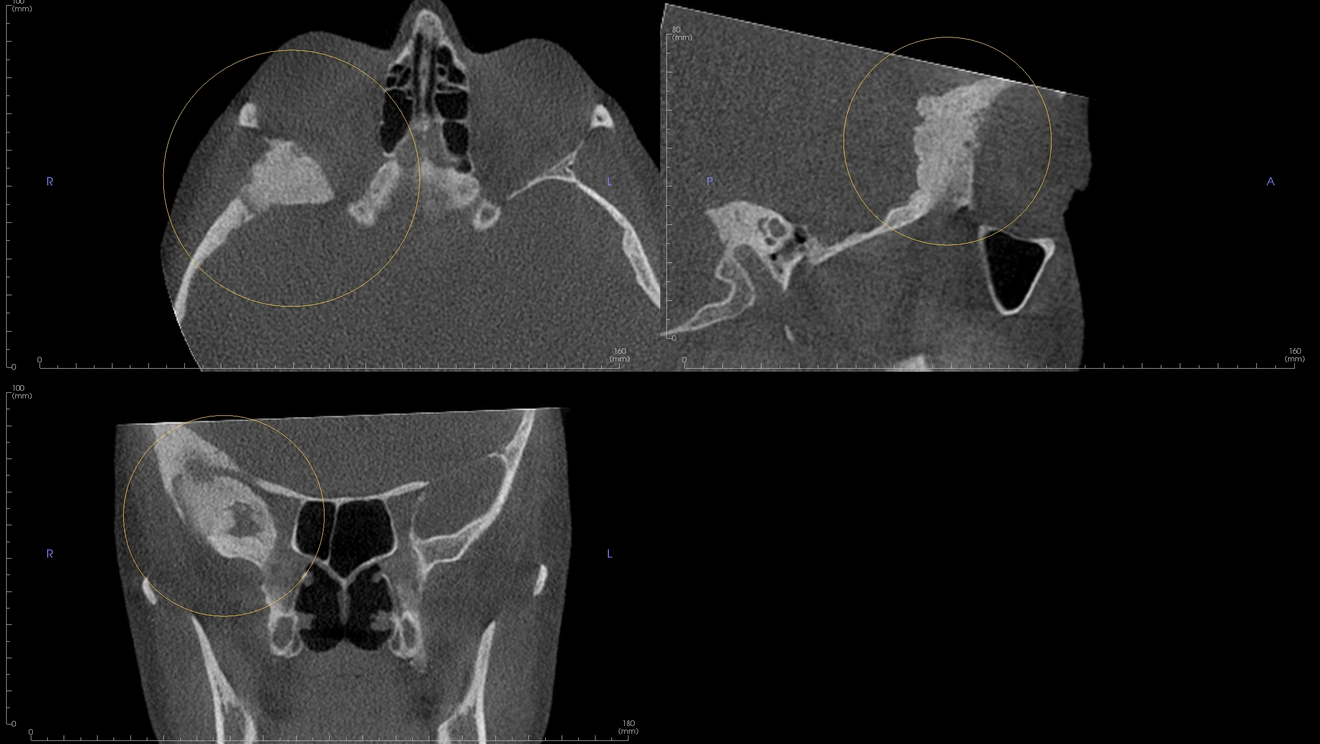
De-identified scan detail.
Registering is easy, free, and secure.
As low as $89 per report, authored by an oral and maxillofacial radiologist. No subscriptions, no minimums.
