43-year-old female
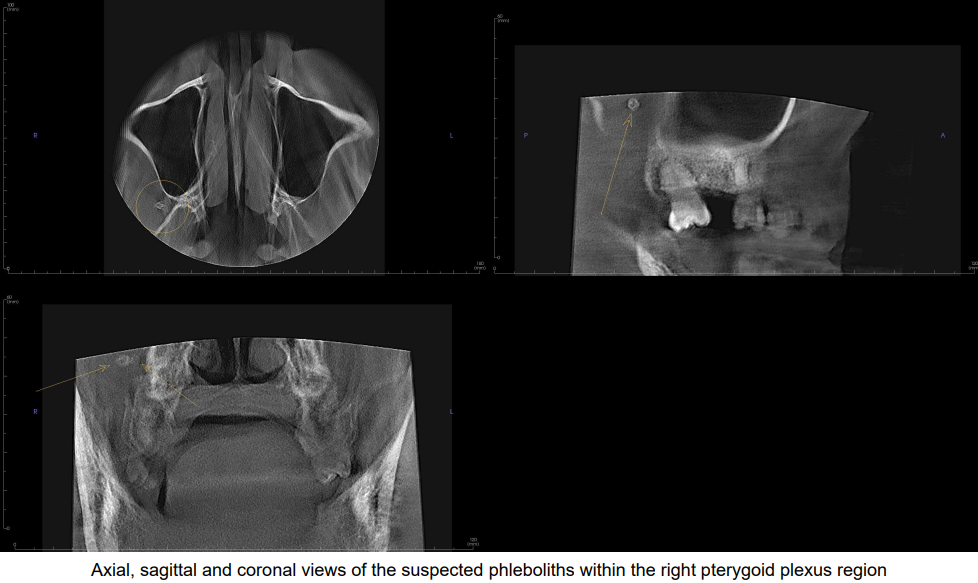
A 43-year-old female with no significant medical history was referred for evaluation of tooth #13. During review, the radiologist identified a cluster of target-shaped lesions within the soft tissue of the right pterygoid plexus region, strongly suggestive of phleboliths, which may indicate the presence of vascular pathology.
Referral for additional imaging with improved soft tissue and vascular contrast was recommended for further evaluation. The radiologist noted that this was a medium field of view study, which is often considered lower risk because fewer anatomic structures are visualized, yet the case demonstrates that significant incidental findings can still occur in non-tooth-bearing regions even at this field of view.
De-identified scan detail.
Registering is easy, free, and secure.
As low as $89 per report, authored by an oral and maxillofacial radiologist. No subscriptions, no minimums.
