17-year-old female
A 17-year-old female was referred for routine post-orthodontic imaging. The patient was asymptomatic with no significant medical history. The radiologist noted this case as particularly interesting because, although the initial abnormality appeared limited to the sphenoid region, systematic review of the full CBCT volume revealed an extensive skull base lesion with widespread craniofacial involvement extending into the optic canal and vidian canal. Referral for multidisciplinary evaluation and advanced imaging was recommended despite the absence of symptoms, as extensive skull base involvement may have important clinical implications.
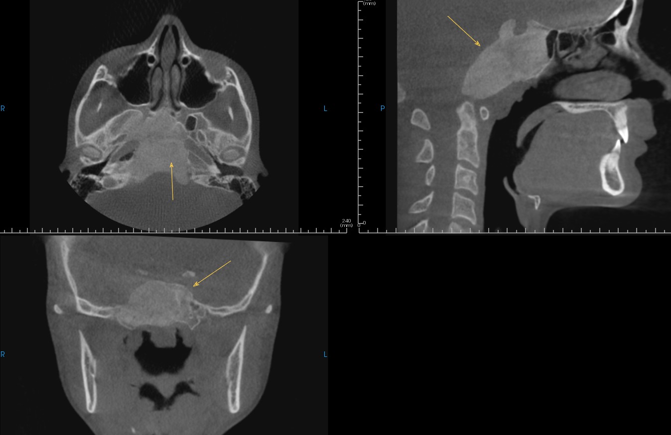
De-identified scan detail.
Registering is easy, free, and secure.
As low as $89 per report, authored by an oral and maxillofacial radiologist. No subscriptions, no minimums.
