
Image Portfolios visually communicate your imaging goals, laying the foundation for patient communication and treatment planning.

A BeamReaders Image Portfolio translates the 3D volume into clear and organized image sets, allowing you to communicate with the patient in a way that's easily understood, custom tuned for your study or treatment purpose.

We work with you to ensure the portfolio gives you what you need to prepare and communicate your treatment plan. Whether you need specific measurements or image sets, our Image Portfolios faciliate optimum planning with accurate scaling.

Let our team of imaging technicians free you and your staff from the time-intensive tasks of image and tracing prep with our extensive library of cephalometric tracings. We can also provide the output in DAZ format for your Dolphin timelines.
Simple Pricing
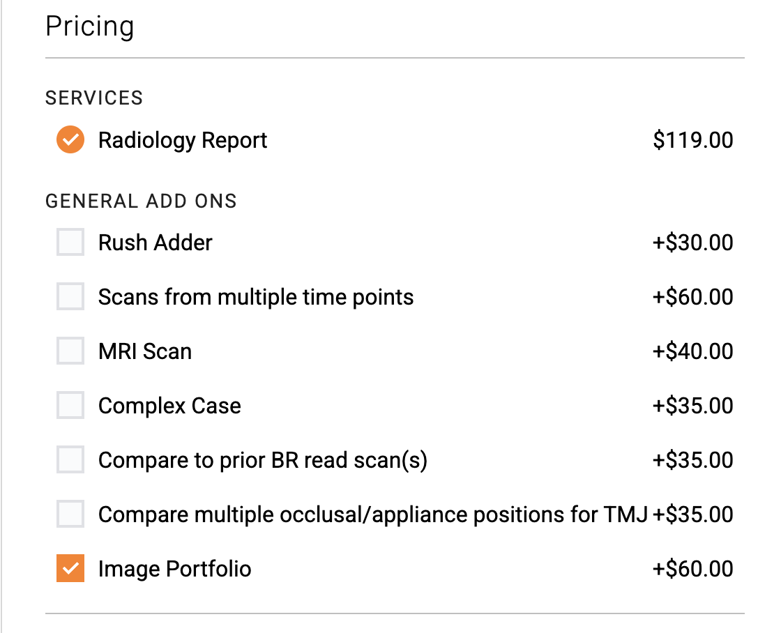
Our Image Portfolios can be ordered individually from a patient profile, or as an optional add-on to a BeamReaders Radiology Report at the time of request.


SAMPLE BeamReaders image portfolios
How to order
Follow these two simple steps inside the VoxDental platform to order an image portfolio without a Radiology Report.
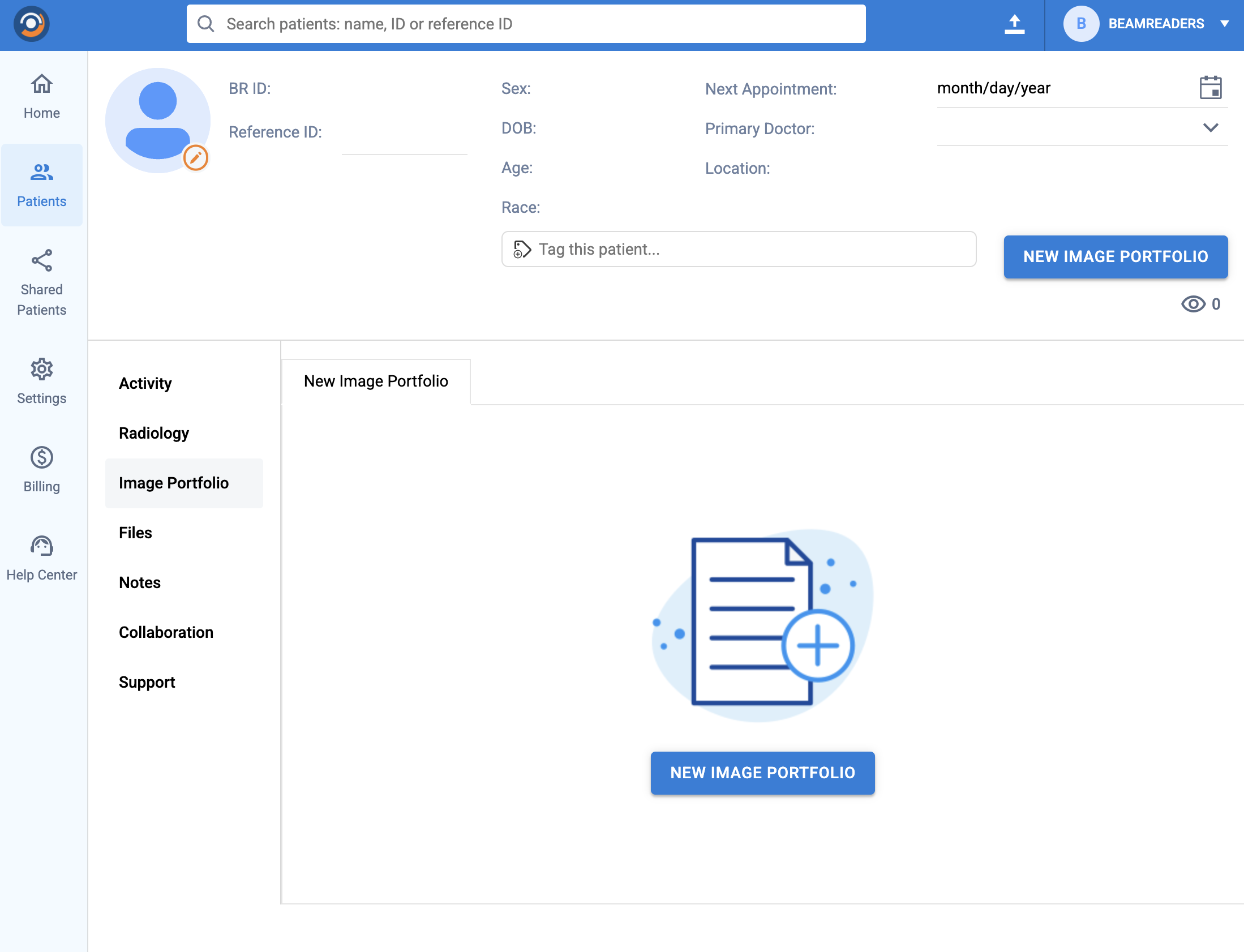
Step 1
Step 2
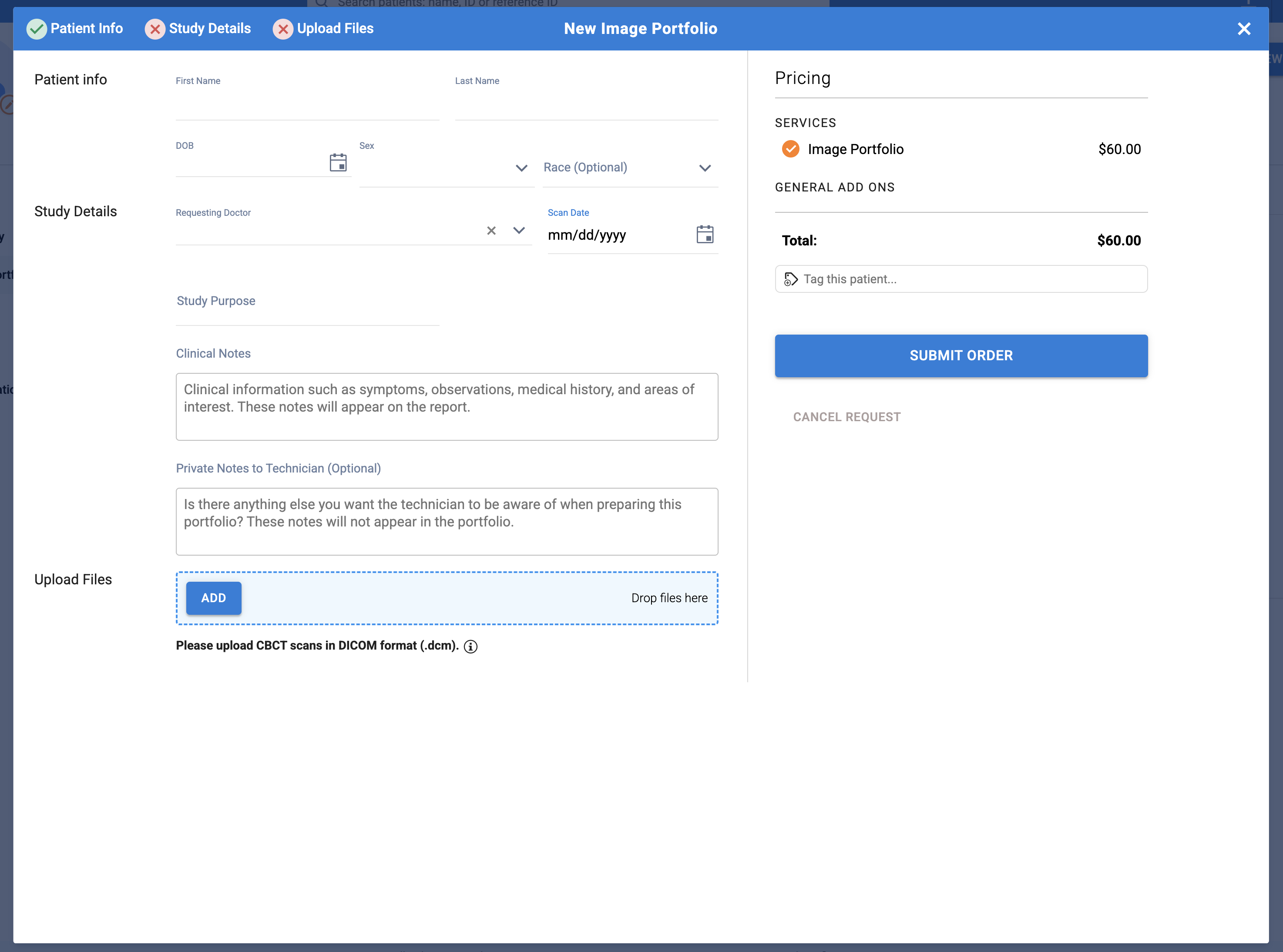
Simply select the Image Portfolio add-on when you order.
One box is all you need
Create your free account and get started with BeamReaders today.